I am doing a project for this quarter, checking to see what-all grows in my home compost. Today the younger stidkid helped me out, taking photographs for me of the various spread plates I have going from a dilution series I started earlier this month. He also helped count the colony forming units (CFUs) for me. He did not have to touch anything “ooky”, and no microorganisms were harmed in the process of our looking at them today. Still, it was pretty cool.
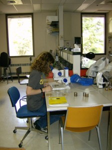
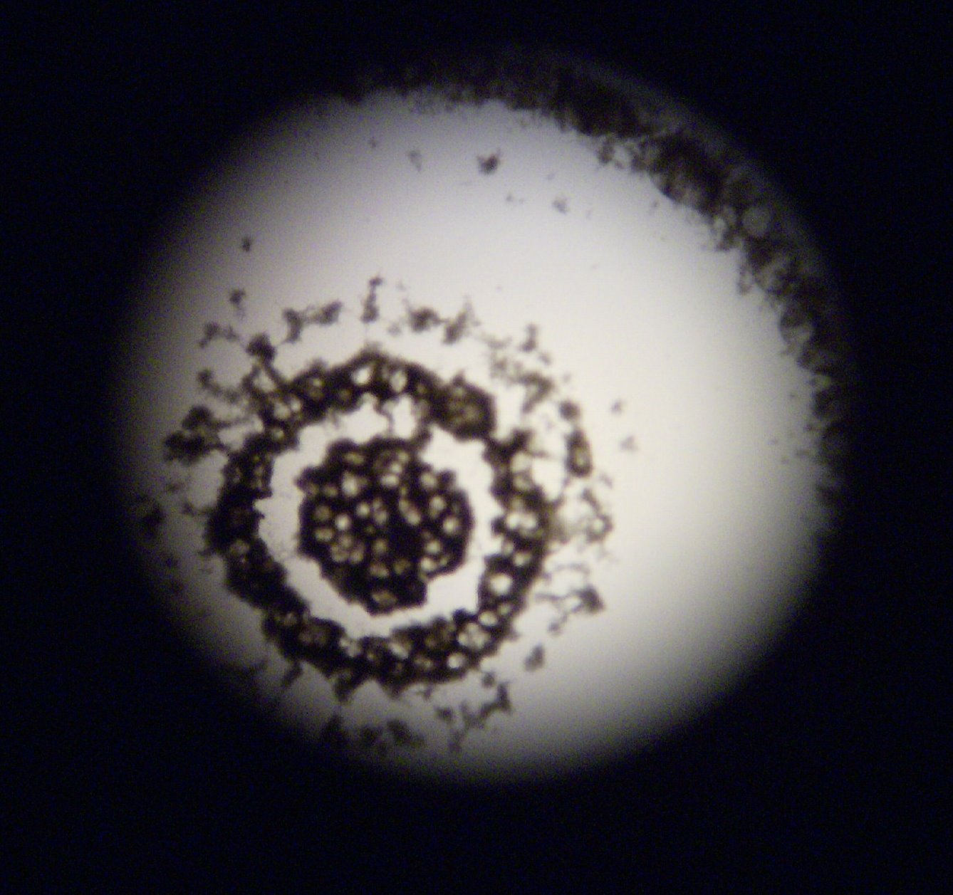
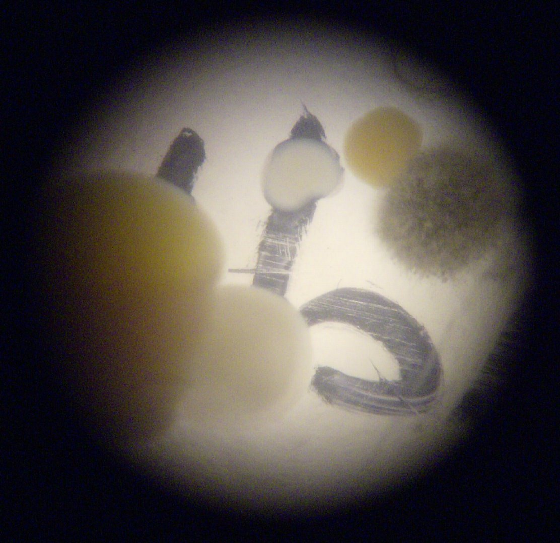
Here he is, looking through a dissecting microscope at some of the plates. The two pics that follow show a “bullseye” fungus (I think) from the top of the pile and a “collection” (bacteria, fungus and possibly yeast) from the middle of the pile are at the setting that is “32” — I think that is total magnification on this microscope (not needing to add in the additional magnification from the eyepiece), but there wasn’t anyone else around to confirm this.
I hope to get more information on this later this week.
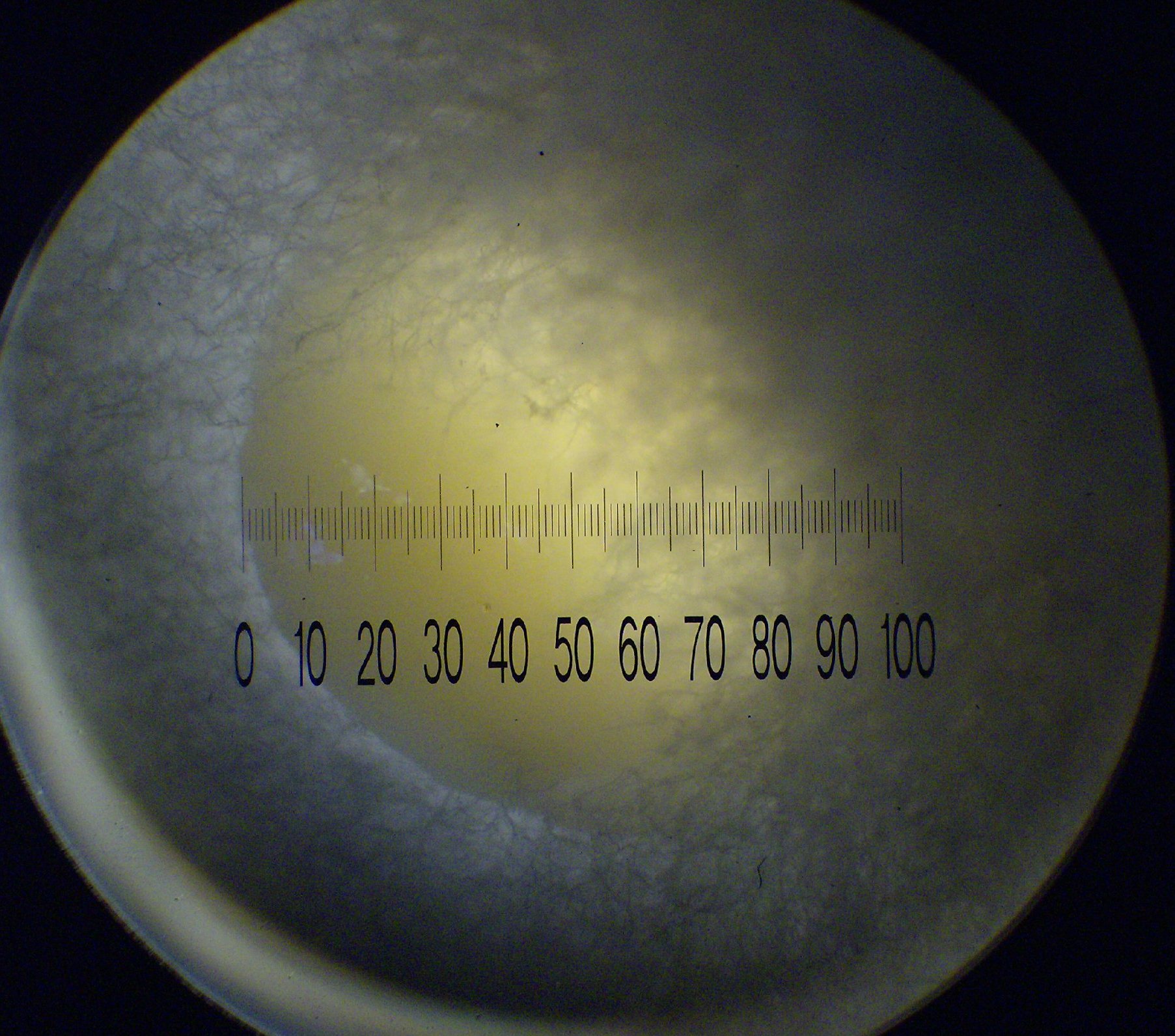
The final pic is through a compound microscope (I think that’s the right term), on the lowest setting which is magnification of 40. You can see a beautiful egg-yolk colored bacterium growing nestled inside a filamentous fungus of some sort. Very cool stuff. I only had a regular ruler to check scale with, but I believe the bacterial colony is about 4mm across, and the fungal colony is at least 10 mm, based on their relative sizes in this picture. The filaments of the fungus are about 1/10 mm across I think.
This was like “take your kid to work day” he got to see where I am two days a week, and participate in the research I am doing. All of a sudden, he is energized and interested in what I am studying. He might not become a microbiologist, but then neither will I. But we will both have a better appreciation for what the real ones do.
Leave a Reply